Parts of the Eye: Understanding the Anatomy and Key Structures
Imagine you’re trying to read the fine print on a product label, only to realize how much you rely on your eyes’ remarkable ability to adjust, focus, and process information. In these moments, you might wonder: what are the parts of the eye that make this all possible? From the delicate lens to the resilient cornea, each structure of the eye has a specific duty, and together they enable the anatomy of eye to function optimally. Understanding the different parts of the eye can help you not only appreciate your vision but also recognize the warning signs when something isn’t working as it should.
As someone who wants to keep their eyesight sharp for years to come, familiarizing yourself with the vital structures of the eye is crucial. Learning about the anatomy of eye, the main components, and how they interact offers valuable insight into vision and eye health. In this guide, we’ll break down each essential piece, so you’ll know what’s happening behind the scenes every time you blink, focus, or take in the world around you.
What Are the Main Parts of the Eye?
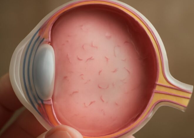
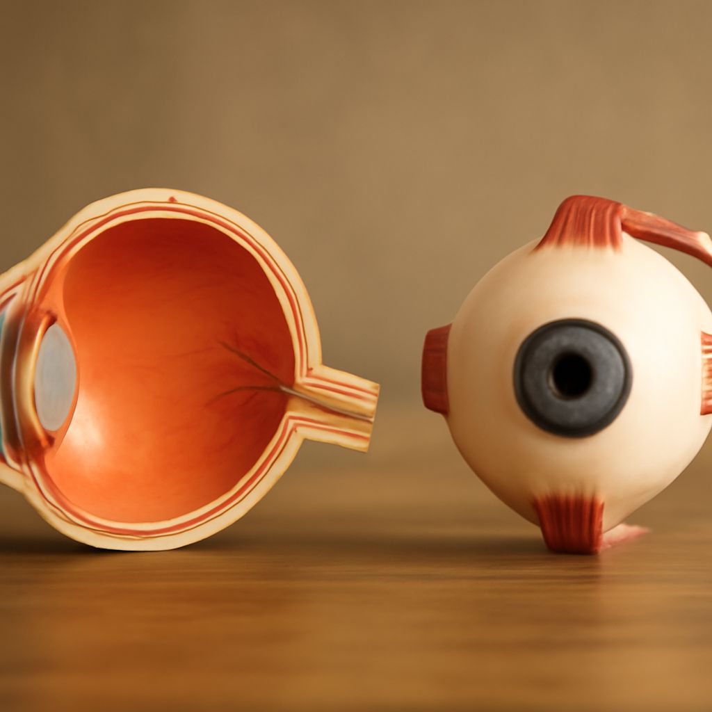
The human eye is a complex organ comprised of several critical parts, each playing a unique role in vision. When discussing the major parts of the eye, we typically focus on the cornea, the iris, the lens, the retina, and the optic nerve. These are the foundational elements that enable us to see clearly and respond to the environment.
The cornea acts as the eye’s transparent, outermost layer, serving as the first barrier and main point of light entry. As light passes through the corneal surface, it is bent or refracted, directing the rays neatly toward the lens. The iris, which gives your eye its distinctive color, works like a camera’s aperture by controlling the amount of light entering through the pupil, its central opening. Adjustments in the iris and pupil diameter allow the eye to adapt to various lighting conditions.
Just behind the pupil sits the lens, which changes shape to focus incoming light onto the retina. This flexibility—commonly called accommodation—enables us to switch from viewing distant objects to focusing on something close up. The retina is the sensitive layer at the back of the eye packed with photoreceptor cells (rods and cones) that capture light and begin the process of visual perception. Finally, the optic nerve transmits visual information from the retina to the brain, where these signals become the images we recognize and understand.
While these are the headline components, other crucial anatomical features, such as the sclera (the white part), conjunctiva, aqueous and vitreous humor, also support the eye’s structure and function. All these main eye parts come together seamlessly, ensuring both protection and performance.
How Does the Anatomy of Eye Support Vision?
The structure and design of the eye’s anatomical elements are finely tuned to provide optimal visual performance. Each component’s arrangement and interaction allow for precise light transmission, focusing, and conversion to neural signals.
Light first encounters the corneal tissue, which starts the bending process necessary for accurate vision. The passage through the aqueous humor—the fluid between the cornea and lens—further directs and nourishes these tissues. After passing the pupil, light reaches the lens apparatus. Through the process of accommodation, the lens alters its curvature depending on whether you’re looking at something near or far. This focusing mechanism is essential for sharp vision and is supported by the flexible zonular fibers and ciliary muscles.
The focused light then strikes the retinal surface, where millions of photoreceptors respond to varying light intensities and color wavelengths. Rod cells assist with night vision and detecting movement, while cone cells are concentrated for sharp, detailed, and color vision. These signals are then bundled and delivered by the optic nerve bundle to the brain’s visual cortex, where interpretation and processing occur.
An efficient anatomy of the eye guarantees that visual data is captured, adjusted, and sent with minimal distortion. When the eye’s structure is altered—by injury, disease, or age—vision may become blurred or impaired. Understanding how this dynamic anatomy underpins vision can help you appreciate the value of routine eye examinations and preventive care.
Which Structures of the Eye Are Most Vital?
While every feature of the eye plays a part, some structures of the eye are particularly essential to maintaining vision quality and eye health. The corneal layer is not only the primary refracting surface but also a crucial barrier against dust, pathogens, and injury. Damage or disease affecting this area, like keratitis or corneal abrasion, can lead to immediate discomfort and vision loss if untreated.
The retinal zone stands out as the critical layer for processing light and transforming it into signals the brain can interpret. Disorders impacting the retina—such as macular degeneration or diabetic retinopathy—often result in significant, sometimes irreversible, vision changes. The optic nerve fibers are equally indispensable; they serve as the information highway connecting the eye to the brain. Conditions like glaucoma target this nerve, potentially causing permanent vision loss if pressure isn’t controlled.
Other supporting parts, such as the lens tissue and the various eye chambers (filled with aqueous and vitreous fluids), maintain the internal shape and clarity of the eye. Each of these vital structures operates together, forming a cohesive network that enables seamless and efficient sight.
Safeguarding these key anatomical features is essential, as their impairment can compromise the entire visual system. Early detection of structural changes ensures that appropriate treatment or intervention can preserve long-term vision.
Why Do the Different Parts of the Eye Matter for Eye Health?
Every unique component among the different parts of the eye contributes to your overall ocular health and functional vision. If one element, such as the corneal surface, suffers damage, your eye’s ability to focus and protect itself diminishes significantly. Maintaining each anatomical region’s health is central to preventing common issues like dry eye, infections, or refractive errors.
For example, clouding of the lens—known as cataract formation—can hinder your vision clarity, requiring surgical removal and replacement. Problems in the retinal area may manifest as dark spots, flashes, or loss of central vision. Since each different segment of the eye is interconnected, a problem in one area often impacts the rest, underlining the need for comprehensive eye care.
Regular checkups help identify early signs of deterioration in these areas, ensuring prompt intervention. Protecting the eye from external hazards, such as excessive sun exposure or trauma, supports the health of these varied anatomical features.
By recognizing the significance of every single eye part, from the iris to the optic nerve, you are better equipped to preserve vision and understand the importance of proactive, preventive eye health strategies.
Key takeaways: The human eye is a sophisticated system of interconnected structures, each vital for clear, healthy vision. Understanding how the various components support vision empowers you to maintain eye health and seek timely care if changes arise.